Imagine a world without sound. No laughter, no music, no whispered secrets. Our ability to perceive this rich auditory landscape, from the faintest rustle of leaves to the roar of a jet engine, hinges on an extraordinary feat of biological engineering deep within your inner ear. Specifically, it's the intricate dance, the vital interaction with Organ of Corti and hair cells, that transforms mere vibrations into the symphony of life your brain understands. This isn't just about hearing; it's about interpreting pitch, volume, and the unique timbre of every sound around you.
This remarkable sensory structure, nestled within the snail-shaped cochlea, acts as your personal sound processor, translating mechanical energy into the precise electrochemical signals that make sense to your brain. It's a complex, delicate system, and understanding how it works is key to appreciating both the marvel of hearing and the challenges when it falters.
At a Glance: Your Inner Ear's Sound System
- The Organ of Corti is your inner ear's primary hearing sensor, converting sound vibrations into brain signals.
- Hair Cells are the stars of the show:
- Inner Hair Cells (IHCs) are the main messengers, sending auditory signals to the brain.
- Outer Hair Cells (OHCs) act as amplifiers and modulators, boosting soft sounds and sharpening frequency detection.
- The Basilar Membrane vibrates in response to sound, acting as a stage for the Organ of Corti.
- Stereocilia, tiny hair-like projections on the hair cells, bend when stimulated, opening channels for electrical signals.
- Endolymph, a potassium-rich fluid, fuels the electrical changes within the hair cells.
- Damage to hair cells is the most common cause of permanent hearing loss, as these cells don't regenerate in mammals.
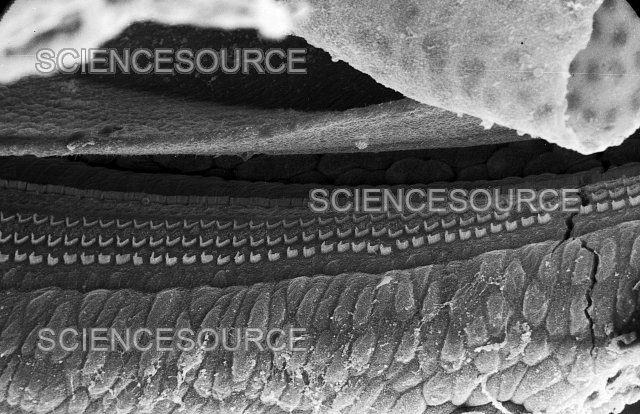
The Inner Ear's Maestro: Unpacking the Organ of Corti
Deep within the temporal bone of your skull lies the cochlea, a tiny, spiraled chamber that houses one of the most sophisticated sensory organs known: the Organ of Corti. Often called the "spiral organ," it's a marvel of precision, designed to capture the subtlest whispers and the loudest roars, translating them into meaningful neural code.
Think of the Organ of Corti as the central processing unit for sound, meticulously arranged along the length of the cochlear duct. This duct, filled with a unique fluid called endolymph, provides the perfect chemical environment for electrical signaling, thanks to its high concentration of potassium ions. This isn't just a static structure; it's a dynamic stage where the mechanical energy of sound gets its first, most crucial transformation.
Meet the Superstars: Inner and Outer Hair Cells
At the heart of the Organ of Corti's function are two distinct populations of sensory cells: the inner hair cells (IHCs) and the outer hair cells (OHCs). While both are vital, they play surprisingly different, yet complementary, roles in how you perceive sound.
Inner Hair Cells: The Primary Messengers
Picture a single, elegant row of about 3,500 cells – these are your inner hair cells. These are the primary transducers of sound. When stimulated, they’re responsible for sending the vast majority of auditory information directly to your brain via the auditory nerve. Each IHC is a dedicated reporter, connecting to roughly ten nerve endings, ensuring that the details of sound are meticulously relayed.
When sound waves ultimately cause these cells to activate, they don't just send a generic "sound received" message. Instead, they encode the intricate characteristics of the sound – its frequency, intensity, and timing – providing the brain with the raw data it needs to construct a rich auditory perception. Without functional inner hair cells, true hearing as we know it simply wouldn't exist.
Outer Hair Cells: The Amplifiers and Modulators
Arranged in three parallel rows, numbering around 12,000 cells, are the outer hair cells. These cells are the unsung heroes of sensitivity and clarity. While they send some signals to the brain, their primary function isn't about transmitting the main auditory message but rather about enhancing it.
Outer hair cells possess a unique superpower called electromotility. This allows them to rapidly change their length, contracting and expanding in response to electrical signals. This rapid movement, largely mediated by a specialized protein called prestin, creates a mechanical amplification within the cochlea. Essentially, OHCs physically boost the vibrations of the surrounding structures, making soft sounds much easier to detect and significantly sharpening the ear's ability to distinguish between very similar frequencies.
Without this amplification, quiet conversations would be muffled, and the subtle nuances that differentiate musical notes or human voices would be lost. The outer hair cells refine your hearing, turning a dull signal into a vibrant, clear perception.
The Architectural Stage: Supporting Structures for Sound
The hair cells don't operate in a vacuum. They are part of a meticulously designed micro-environment, supported by several key structures that are crucial for their function.
The Basilar Membrane: Your Personal Soundboard
Underpinning the entire Organ of Corti is the basilar membrane, a flexible, fibrous ribbon that stretches the length of the cochlea. Imagine it as a finely tuned instrument string, but one that responds differently along its length. It forms the floor of the cochlear duct, separating it from the lower scala tympani.
When sound energy enters the cochlea, it sets the basilar membrane into a ripple-like motion, creating what's known as a "traveling wave." Crucially, different frequencies of sound cause this membrane to ripple most intensely at specific locations along its length. This phenomenon, called tonotopic organization, means that high frequencies cause maximum ripple near the base of the cochlea, while low frequencies do so closer to the apex. This spatially organized vibration pattern is the first step in the ear's ability to differentiate pitch. You can visualize this intricate structure with the basilar membrane schematic.
The Tectorial Membrane: The Gelatinous Lid
Suspended directly above the hair cells, acting like a soft, gelatinous roof, is the tectorial membrane. This structure is vital for creating the mechanical force that stimulates the hair cells. The stereocilia (the hair-like projections) of the hair cells are either embedded in or come into contact with this membrane. Its relative stability, compared to the vibrating basilar membrane, is what generates the critical shearing motion that drives hearing.
Endolymph: The Powerhouse Fluid
The cochlear duct, where the Organ of Corti resides, is filled with a unique fluid called endolymph. Unlike other body fluids, endolymph is extraordinarily rich in potassium ions and has a positive electrical potential. This creates a powerful electrochemical gradient across the hair cell membranes. This high potassium concentration is absolutely essential for the rapid depolarization of hair cells, making the swift conversion of mechanical motion into electrical signals possible. It's the battery that powers your hearing.
Supporting Cells: The Structural Backbone
Rounding out the Organ of Corti are various supporting cells. These aren't directly involved in transducing sound but are indispensable for maintaining the structural integrity of the organ. They hold the delicate hair cells in place, provide metabolic support, and contribute to the overall specialized environment necessary for optimal hearing function.
The Symphony of Sound: Step-by-Step Interaction
So, how do all these parts work together to transform a simple sound wave into something you can understand? It’s a beautifully choreographed sequence of events:
- Sound Waves In, Fluid Waves Out: The journey begins with sound waves vibrating the eardrum and the tiny bones of your middle ear (ossicles). This mechanical energy is then transmitted to the fluid within the cochlea, generating fluid movement.
- The Traveling Wave: This fluid movement creates a propagating ripple, a "traveling wave," along the length of the basilar membrane. As we discussed, the point on the basilar membrane where this wave reaches its peak is determined by the sound's frequency – its pitch.
- Up and Down, Creating a Shear: As the basilar membrane vibrates, it moves the entire Organ of Corti up and down. Because the tectorial membrane above remains relatively stable, this vertical movement creates a powerful shearing force between the two membranes.
- Stereocilia Bend, Channels Open: This shearing force causes the tiny, stiff hair-like projections atop the hair cells, known as stereocilia, to bend. Crucially, these stereocilia are connected by ultrafine filaments called tip links. When the stereocilia bend, these tip links pull open specialized cation (ion) channels located at their tips.
- Potassium In, Cell Depolarizes: With the channels open, there's a rapid, sudden influx of positively charged potassium ions from the potassium-rich endolymph into the hair cell. This surge of positive charge rapidly changes the electrical potential across the hair cell's membrane, a process called depolarization.
- Neurotransmitter Release, Signal to Brain: This electrical depolarization triggers the release of a chemical messenger, a neurotransmitter called glutamate, from the base of the hair cell. This release is particularly prominent from the inner hair cells, which are the primary communicators with the brain.
- Auditory Nerve Activation: Glutamate then binds to receptors on the adjacent auditory nerve fibers, initiating electrical impulses (action potentials) in these nerve cells. These impulses are then faithfully transmitted along the auditory nerve, making their way through various relay stations (spiral ganglion, inferior colliculus, medial geniculate nucleus) until they reach the auditory cortex in your brain, where they are interpreted as sound.
The OHC "Superpower": Amplification and Sharpening
While this process describes how sound becomes an electrical signal, the outer hair cells add a critical layer of refinement. As they receive subtle electrical feedback, their electromotility kicks in. They actively contract and expand, physically boosting the basilar membrane's movement precisely at the frequency being stimulated.
This active process significantly amplifies soft sounds, making them audible, and it sharpens the tuning of the basilar membrane, allowing for incredibly precise discrimination between frequencies. This OHC amplification is so powerful that it can actually generate faint sounds that travel back out of your ear canal – these are called otoacoustic emissions, and measuring them is a standard, non-invasive way to screen for hearing in newborns.
Why This Interaction Matters: More Than Just "Hearing"
The complex interaction within the Organ of Corti and its hair cells isn't just about detecting the presence of sound. It's about translating the full richness of our auditory world.
- Pitch Perception: The tonotopic organization of the basilar membrane, where different frequencies vibrate different regions, is the foundation of your ability to discern high notes from low notes.
- Volume (Loudness) Perception: Louder sounds cause greater displacement of the basilar membrane, leading to more intense bending of stereocilia, a larger influx of potassium, and consequently, a greater release of neurotransmitter and higher firing rates in the auditory nerve.
- Timbre (Sound Quality) Perception: The unique quality of a sound – what makes a flute sound different from a violin playing the same note – is due to its complex mix of frequencies (harmonics). The precise frequency tuning provided by the Organ of Corti, especially with OHC amplification, allows your brain to analyze these complex sound profiles and distinguish their unique qualities.
This intricate interplay of mechanics, fluid dynamics, and electrochemistry ensures that the sounds reaching your ears are not just registered, but meticulously analyzed and conveyed to the brain for complete comprehension.
When the Symphony Falters: Understanding Hearing Loss
Given the delicate nature and precision of the Organ of Corti and its hair cells, it's perhaps not surprising that they are vulnerable to damage. In fact, damage to these sensory hair cells is the most common cause of permanent sensorineural hearing loss. What makes this particularly impactful is a crucial biological fact: mammalian hair cells do not regenerate. Once they are damaged or die, they are gone forever, making the resulting hearing loss irreversible.
Understanding the primary culprits behind this damage is essential for prevention.
Acoustic Trauma: The Loud Noise Culprit
One of the most insidious threats to your hearing comes from acoustic trauma, prolonged or intense exposure to loud sounds. While the occasional loud concert might seem harmless, sustained exposure to sounds over 85 decibels (the equivalent of heavy city traffic) can cause physical damage or outright destruction of the delicate hair cells and their stereocilia.
Imagine a violent earthquake shaking the basilar membrane. The extreme vibrations can shear off stereocilia, disrupt the intricate cellular structure, or even completely obliterate the hair cells. This often leads to immediate or gradual hearing loss, sometimes accompanied by tinnitus (ringing in the ears). Protecting your ears from excessive noise is paramount.
Age-Related Loss (Presbycusis): The Natural Decline
As we age, our bodies undergo natural wear and tear, and the auditory system is no exception. Presbycusis, or age-related hearing loss, is a common condition resulting from the gradual degradation and death of hair cells over a lifetime.
This process typically begins to affect the high-frequency range first, which is primarily processed by hair cells located at the base of the cochlea. You might first notice difficulty understanding speech in noisy environments or struggling to hear high-pitched sounds like birdsongs or certain consonants. It's a natural, though often frustrating, part of aging for many individuals.
Ototoxic Medications: Chemical Attacks
Certain medications, while vital for treating various diseases, can unfortunately have toxic effects on the auditory system, particularly on hair cells. These are known as ototoxic medications. Common examples include some chemotherapy agents (like cisplatin) and specific classes of antibiotics (such as aminoglycosides).
These drugs can chemically poison the hair cells, interfering with their metabolic processes or directly leading to their demise. The effects can be dose-dependent and sometimes reversible if caught early, but severe ototoxicity can cause permanent hearing loss. If you are prescribed such medications, your doctor will often monitor your hearing carefully.
Navigating Hearing Challenges: What Happens Next?
Given the irreversibility of hair cell damage, early detection and appropriate management of hearing loss are crucial. This is particularly true for children, where early intervention can have a profound impact on language development.
When the interaction within the Organ of Corti is compromised, different solutions become necessary:
- Hearing Aids: For many forms of sensorineural hearing loss, especially those involving partial damage to hair cells, hearing aids can be highly effective. These devices amplify sound, helping the remaining healthy hair cells to be stimulated more effectively. They essentially boost the signal that your compromised hair cells can still pick up.
- Cochlear Implants: When hair cell damage is severe or complete, and hearing aids no longer provide sufficient benefit, a cochlear implant may be an option. Unlike hearing aids, which amplify sound, a cochlear implant bypasses the damaged Organ of Corti entirely. It's an electronic device that directly stimulates the auditory nerve, allowing electrical impulses to be sent to the brain, restoring a sense of hearing.
- Sign Language and Other Communication Strategies: For individuals with profound hearing loss, or those who choose alternative paths, learning sign language becomes a powerful tool for communication and community. Other strategies include lip-reading, assistive listening devices, and adapting communication environments.
The Impact of Outer Hair Cell Loss
It's worth noting that the specific type of hair cell damage influences the nature of hearing loss. A significant loss of outer hair cells, for example, primarily leads to two key issues:
- Reduced Amplification for Quiet Sounds: Without the OHCs' active boosting mechanism, soft sounds simply aren't strong enough to effectively stimulate the inner hair cells, making it difficult to hear whispers or the subtle nuances in speech.
- Difficulty Distinguishing Frequencies: The OHCs play a critical role in sharpening the frequency tuning of the basilar membrane. When they are lost, the ear's ability to precisely separate one frequency from another is impaired, leading to a perception of "muddiness" in sound, where distinct pitches blend together. This can make understanding speech in noisy environments particularly challenging, as it becomes harder to separate the voice you want to hear from background noise.
The brain plays a significant role in further processing auditory information, including recognizing and localizing sounds, through parallel streams that begin receiving signals from the auditory nerve. But the quality of those initial signals, generated by the Organ of Corti, is paramount.
Your Blueprint for Hearing Health: Protecting Your Inner Ear
While the inability of mammalian hair cells to regenerate is a stark reality, you have significant power to protect the hearing you have. Understanding the delicate mechanics of the Organ of Corti highlights the importance of preventative measures:
- Mind Your Volume: Be acutely aware of noise levels in your environment. Use hearing protection (earplugs, earmuffs) when exposed to loud machinery, concerts, sporting events, or even prolonged use of headphones. Remember the 85-decibel rule: if you have to shout to be heard over background noise, it's probably too loud.
- Regular Check-ups: Incorporate hearing screenings into your routine medical check-ups, especially as you age or if you have a family history of hearing loss. Early detection can lead to better management and intervention.
- Medication Awareness: Discuss with your doctor any concerns about ototoxic medications. If you must take them, ensure you are monitored for any auditory side effects.
- Healthy Lifestyle: General health practices, such as managing blood pressure, diabetes, and maintaining good cardiovascular health, indirectly support the delicate blood supply and overall function of your inner ear.
- Give Your Ears a Break: If you're in a noisy environment, step away for a few minutes every hour to give your hair cells a chance to recover.
Common Questions About Your Inner Ear's Sound Factory
What is the main function of the Organ of Corti?
The Organ of Corti's main function is to convert the mechanical energy of sound vibrations into electrochemical signals that the brain can interpret. This allows us to perceive pitch, volume, and timbre.
How do inner and outer hair cells differ in their roles?
Inner hair cells (IHCs) are the primary transducers, sending the main auditory signal to the brain. Outer hair cells (OHCs) primarily function as amplifiers and modulators, rapidly changing length to boost soft sounds and sharpen frequency tuning.
Can damaged hair cells regenerate in humans?
No, mammalian hair cells (including human hair cells) do not regenerate. Once damaged or destroyed, the resulting sensorineural hearing loss is permanent.
What are otoacoustic emissions, and what do they indicate?
Otoacoustic emissions (OAEs) are faint sounds generated by the active amplification process of the outer hair cells that travel back out of the ear canal. Their presence indicates healthy outer hair cell function and are commonly used in newborn hearing screenings.
The Unseen Marvel: A Deeper Appreciation for Your Hearing
The intricate interaction with the Organ of Corti and hair cells is not just a biological mechanism; it's the very foundation of your auditory world. It’s a testament to the sophistication of the human body, transforming unseen air vibrations into meaningful experiences – the comfort of a loved one's voice, the energy of your favorite music, the warning of an approaching car.
Understanding this vital process underscores the importance of protecting your hearing. Treat your ears with the care they deserve, and you can continue to enjoy the rich, diverse symphony of life for years to come.